Bone Cross Section Diagram / Instant Anatomy - Lower Limb - Areas/Organs - Thigh ... / Compact bone is the outer layer and the spongy bone forms the inner layer.
Bone Cross Section Diagram / Instant Anatomy - Lower Limb - Areas/Organs - Thigh ... / Compact bone is the outer layer and the spongy bone forms the inner layer.. Diagram with articular cartilage, marrow, medullary cavity and periosteum. Spinal cord spinal column anatomy information myvmc. Vector illustration scheme of bone cross section. They are similar to the topographic profiles that you created in the topographic maps chapter, but they also show the rock types and geologic structures. Vector illustration scheme of bone cross section.
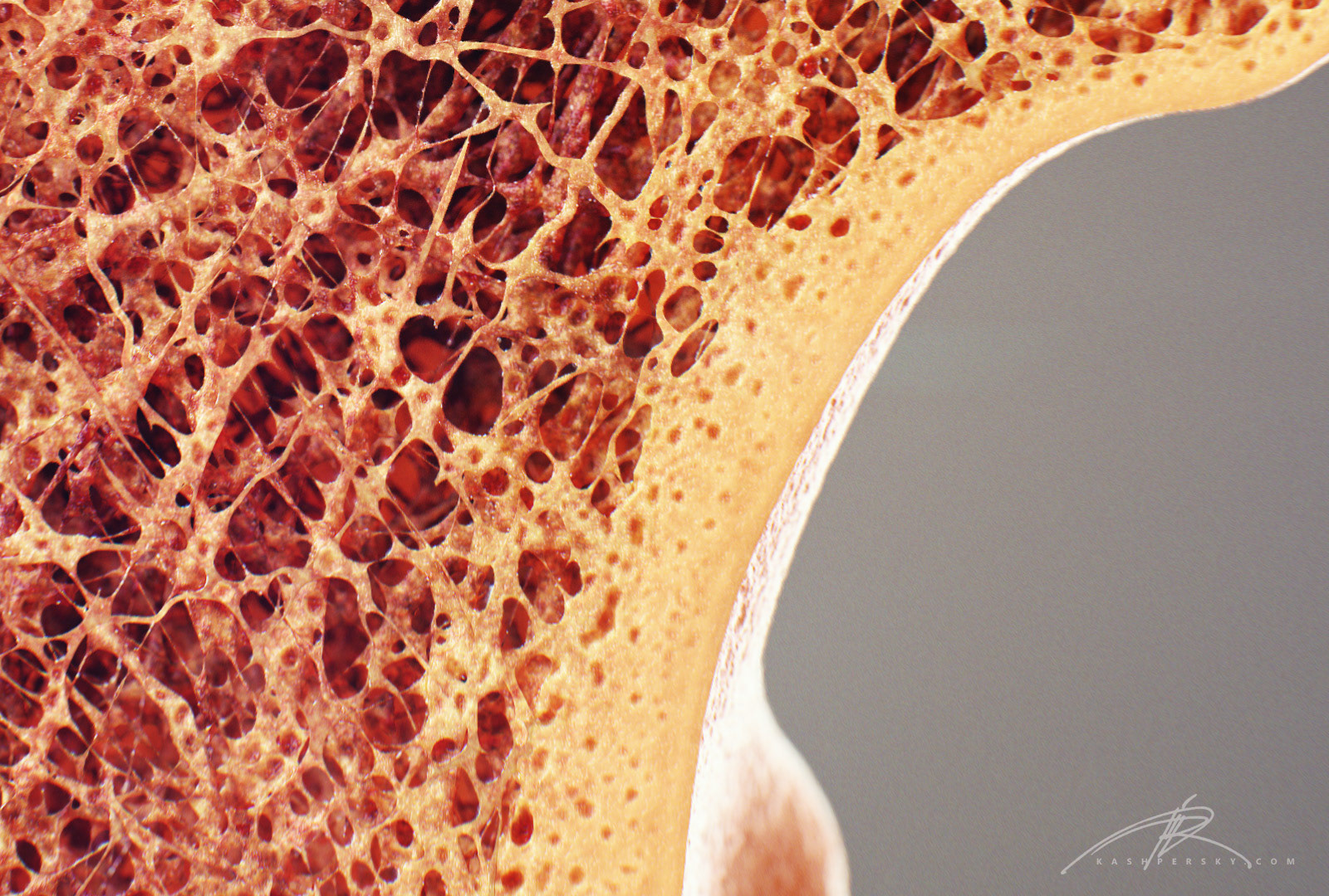
Diagram with articular cartilage, marrow, medullary cavity and periosteum. There are trabeculae in spongy bone which gives its sponge like appearance. 850 x 1270 png 173kb. The vascular section contains blood vessels that supply the bone with nutrients and transport blood stem cells and formed mature blood cells this article has clear diagrams/pictoral representations which i would like to use for teaching purposes. As shown in figure 2.
Vector illustration scheme of bone cross section. (b) in this micrograph of the osteon, you can clearly see the concentric lamellae and central canals. Schematic drawing of a longitudinal section through a. Cross section of the human retina. Jump to navigation jump to search. Diagram of a cross section of the coiled cochlea. Spinal cord spinal column anatomy information myvmc. Medically reviewed by the healthline medical network — written by the healthline editorial team — updated on january 20, 2018. As shown in figure 2. Crosssection cutaway diagram dry cell battery. Compact bone is the outer layer and the spongy bone forms the inner layer. Spongy bone and compact bone. They build the entire picture, improve your understanding, consolidate the information and facilitate recall.
Diagram with articular cartilage, marrow, medullary cavity and periosteum. Two prominent grooves or sulci run along its length. As shown in figure 2. Explaned distal and proximal epiphysis. (b) in this micrograph of the osteon, you can clearly see the concentric lamellae and central canals.
Newt Studios - Bone Cross Section from pro2-bar-s3-cdn-cf1.myportfolio.com As shown in figure 2. Fermur bone with labels and diagram. 850 x 1270 png 173kb. Spinal cord spinal column anatomy information myvmc. 512 x 512 jpeg 27kb. Diagram of a cross section of the coiled cochlea. Spongy bone and compact bone. Two prominent grooves or sulci run along its length.
Vector illustration scheme of bone cross section.
Vector illustration scheme of bone cross section. They build the entire picture, improve your understanding, consolidate the information and facilitate recall. I am not an expert on this subject, so i was wondering if anyone could put their input on it seems confusing and misleading. They are similar to the topographic profiles that you created in the topographic maps chapter, but they also show the rock types and geologic structures. It consists of two layers; For example, to read this diagram literally, since the cartilage can be seen inside the cutaway section of bone, it. Healthy tooth diagram isolated on white background vector. This is a short tutorial using blender 2.8 that shows how to create a bone cross section and using images to create the textures. Explaned distal and proximal epiphysis. Diagram with articular cartilage, marrow, spongy bone, medullary cavity, endosteum, diaphysis, and periosteum. Fermur bone with labels and diagram. (b) in this micrograph of the osteon, you can clearly see the concentric lamellae and central canals. Volcano cross section diagram drawing high.
Please will you consider sharing with me? Medically reviewed by the healthline medical network — written by the healthline editorial team — updated on january 20, 2018. It consists of two layers; There are trabeculae in spongy bone which gives its sponge like appearance. Vector illustration scheme of bone cross section.


Posting Komentar
0 Komentar